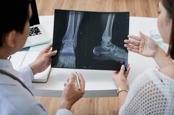
X-Ray Imaging

X-Ray Imaging is a widely used diagnostic tool in medicine, utilizing electromagnetic radiation to create images of the inside of the body. It is primarily used to examine bones, detect abnormalities in the chest, and evaluate various tissues and organs. X-rays pass through the body and are absorbed by different tissues at varying rates. The resulting image, known as a radiograph, helps healthcare professionals diagnose conditions ranging from fractures to infections, tumors, and other internal abnormalities.
Principles of X-Ray Imaging:
- Electromagnetic Radiation: X-rays are a form of high-energy radiation, part of the electromagnetic spectrum. They are able to penetrate the body due to their high energy.
- Absorption: Different tissues in the body absorb X-rays at different rates. Dense structures like bones absorb more X-rays and appear white on the image, while softer tissues (muscles, organs) absorb fewer X-rays and appear darker.
Types of X-Ray Examinations:
Plain X-Ray (Radiograph):
- The most common and straightforward type of X-ray. It is used to examine bones for fractures, dislocations, and joint problems, as well as to assess lung conditions, such as pneumonia or tuberculosis.
Fluoroscopy:
- A type of X-ray that produces real-time moving images. It is often used during procedures like catheter placements, barium swallow studies, or guiding surgical instruments during certain types of surgery.
Mammography:
- A specialized form of X-ray imaging used to examine breast tissue for abnormalities, including tumors or cysts. Mammography is essential for breast cancer screening.
Computed Tomography (CT) Scan:
- A more advanced form of X-ray imaging that provides cross-sectional images (slices) of the body. CT scans can offer more detailed views of internal structures than standard X-rays and are frequently used for diagnosing conditions in the abdomen, chest, and brain.
Indications for X-Ray Imaging:
- Fractures and Dislocations: To detect bone fractures, joint dislocations, and alignment issues.
- Chest Conditions: To diagnose respiratory infections (e.g., pneumonia, tuberculosis), lung cancer, and conditions such as heart failure or pleural effusion.
- Infections and Inflammation: To detect infections in bones (osteomyelitis) or joints (arthritis).
- Tumors: To identify masses or abnormal growths in various organs or tissues.
- Dental Conditions: For evaluating teeth and surrounding bone structures, identifying cavities, or assessing alignment.
Advantages of X-Ray Imaging:
- Non-invasive: X-ray imaging is typically quick and does not require incisions or other invasive procedures.
- Widely Available: X-ray machines are common in hospitals and clinics and can be accessed in emergency and outpatient settings.
- Quick Results: Radiographs can be obtained and analyzed quickly, providing immediate feedback in many cases.
- Cost-effective: Compared to more advanced imaging modalities like MRI or CT, plain X-rays are relatively inexpensive.
Disadvantages of X-Ray Imaging:
- Radiation Exposure: X-rays expose the body to a small amount of ionizing radiation, which, in high doses or repeated exposure, may increase the risk of cancer. However, the radiation dose in a typical X-ray is minimal and is generally considered safe for most patients.
- Limited Soft Tissue Detail: X-rays are most effective at visualizing dense structures like bones. They offer limited information about soft tissues like muscles, tendons, or internal organs, which may require additional imaging techniques (e.g., ultrasound, MRI).
- Pregnancy: Due to the potential risk of radiation to the fetus, X-rays are usually avoided during pregnancy, especially in the first trimester, unless the procedure is essential and the benefits outweigh the risks.
Preparation for an X-Ray Exam:
- Clothing: Patients are often asked to remove clothing from the area being examined and wear a hospital gown to avoid interference with the image.
- Metal Objects: Jewelry, dentures, or other metal items should be removed, as they can block the X-ray beam and distort the image.
- Positioning: The patient may be asked to stand, sit, or lie down in a specific position, depending on the area being examined. In some cases, multiple images from different angles may be taken to provide a complete assessment.
Interpretation of X-Ray Images:
Radiologist
: A radiologist, a doctor specialized in medical imaging, interprets the X-ray images. They look for signs of fractures, infections, tumors, or other abnormalities.- Contrast: Areas that appear white on the X-ray are denser, such as bones, while darker areas represent less dense tissues (e.g., muscles, fat).
- Further Investigation: If abnormalities are detected on an X-ray, further tests or imaging may be required to confirm a diagnosis or evaluate the extent of the condition.
Common Conditions Diagnosed with X-Ray:
- Fractures: Broken bones, including simple fractures or complex fractures involving multiple bone fragments.
- Arthritis: Joint degeneration, bone spurs, and changes in bone density due to arthritis.
- Infections: Pneumonia, tuberculosis, or osteomyelitis.
- Bone Tumors: Primary or metastatic tumors affecting the bones.
- Lung Conditions: Pneumonia, pleural effusion, pulmonary edema, or lung cancer.
Conclusion:
X-ray imaging is an essential tool in modern medicine, offering a fast, cost-effective, and non-invasive method for diagnosing a wide range of conditions, particularly those involving bones and lungs. Although it has limitations in visualizing soft tissue, its use remains critical in both emergency and routine diagnostic settings. The risk of radiation exposure is minimal and generally outweighed by the benefits in diagnosing serious health conditions, and with technological advancements, X-ray imaging continues to improve in precision and safety.


